YOUR BROWSER IS OUT-OF-DATE.
We have detected that you are using an outdated browser. Our service may not work properly for you. We recommend upgrading or switching to another browser.
Date: 28.07.2020 Category: science/research/innovation
An ultra-compact laser, which will facilitate accurate imaging of the retina and earlier detection of eye diseases, has been developed by researchers from the Faculty of Electronics. The results of their research were published in the renowned scientific journal Biomedical Optics Express.

Since 2018, Grzegorz Soboń, PhD, DSc. and his team have been working on new type lasers – so-called optical frequency combs. As part of the project named "Fiber-based mid-infrared frequency combs for laser spectroscopy and environmental monitoring", financed by the Foundation for Polish Science, they have just created a prototype of an ultra-compact laser. - It’s the so-called femtosecond laser, which can be used in the imaging of biological tissues – explains head of the project Grzegorz Soboń, PhD, DSc. - It’ll be used for the in vivo imaging of the retina of the eye, among other things, thus enabling the creation of tools for advanced and early diagnosis of eye diseases.
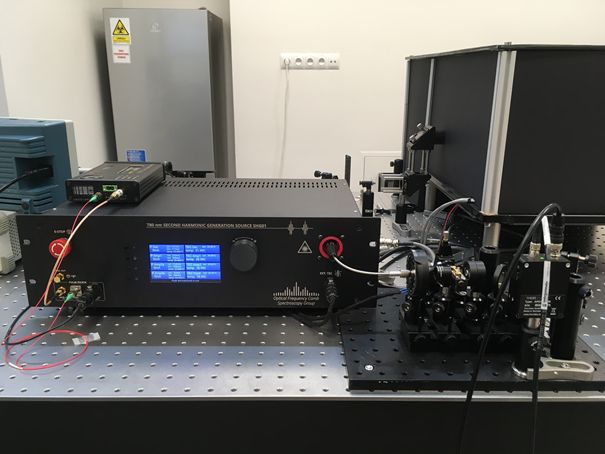
 The prototype, developed at WUST’s Faculty of Electronics, has unique parameters, unattainable by other systems currently available on the market. The laser generates ultra-short, i.e. 60 fs (60×10-15 of a second) pulses with a wavelength of 780 nm (i.e. near the border of the visible and infrared band) and allows the tuning of the pulse repetition frequency (i.e. adjustment of the time interval between successive pulses). Especially the latter feature is crucial for applications in multi-photon microscopy, as it makes it possible to adjust the pulse frequency to specific fluorophores.
– We’ve shown that increasing the interval between pulses while maintaining their duration, enables increasing the intensity of the fluorescent signal of the sample being measured - says Dr Soboń. – This is important in the case of tests of tissues sensitive to damage, such as the human eye, for which high optical power can’t be used.
Our scientists also wanted to simplify the laser design as much as possible. And they succeeded. The prototype does not require any adjustment or calibration and it can be operated by medical personnel, doctors, and biologists. – It is a fibre optical laser, i.e. the light is "trapped" in the optical fibres and leaves them only at the very end of the system, before a two-photon microscope – explains the WUST researcher. – Thanks to its simple design, which uses our "know-how" in the area of amplifying ultra-short laser pulses and non-linear phenomena occurring in optical fibres, the device is also much cheaper to produce than the competitive titanium-sapphire laser – he adds.
 Thanks to the cooperation with the team headed by Professor Maciej Wojtkowski (winner of the FNP, so-called "Polish Nobel Prize” and pioneer in the field of optical coherence tomography of the eye) from the Institute of Physical Chemistry of the Polish Academy of Sciences, the laser from Wrocław was integrated with a two-photon fluorescence microscope built at the IPC.
– We used the laser for the imaging of selected biological tissues ex vivo, such as frog's liver and rat skin, or certain plants – says Dr Grzegorz Soboń. – We’ve shown that with reduced pulse repetition frequency, it’s possible to obtain a proportionally higher fluorescent response, which makes it possible to obtain excellent quality images of tissues, without the risk of damaging them. Importantly, the parameters of radiation generated by the laser also meet the safety requirements for the human application – says the researcher from W4.
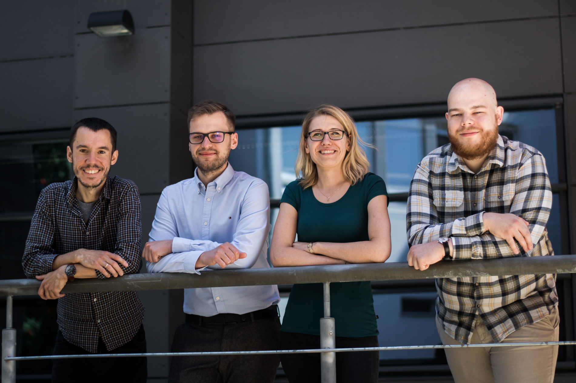
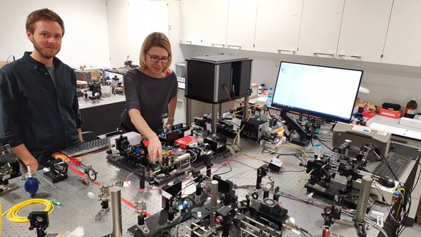
The main designer of the laser is Dorota Stachowiak, PhD, Eng. from the Department of Field Theory, Electronic Systems, and Optoelectronics, while the research into fluorescent microscopy was carried out by Jakub Bogusławski, PhD, Eng., who received his doctorate at WUST and currently works at the Institute of Physical Chemistry of the Polish Academy of Sciences. Aleksander Głuszek and Zbigniew Łaszczych also work in the team.
Our scientists' paper in which they describe the laser’s design and its application in microscopy, has just been published in the renowned scientific journal Biomedical Optics Express.
Our site uses cookies. By continuing to browse the site you agree to our use of cookies in accordance with current browser settings. You can change at any time.